
A new kinematic protocol for evaluating sagittal posture during walking: the DB-Total protocol

Author: Marta Francisca Corrà, Ph.D.
Posture is defined as the position of human body segments and their orientation, and is usually assessed clinically by the physician, with high inter-operator variability [1]. Another more standardized method is instrumental, which allows quantification of postural elements such as stability, morphology and symmetry of musculoskeletal districts, and plantar pressures under static and dynamic conditions [2,3].
The traditional instrumented method for assessing postural stability is the stereophotogrammetric model [4], which evaluates the kinematics of the pelvis, hip, knee, ankle, trunk, and, rarely, head and upper limbs. In particular, the trunk is considered as a single rigid segment that does not provide information about kinematic changes within the spine. Only a few studies have analyzed the multi-segmental trunk in standing position [5].
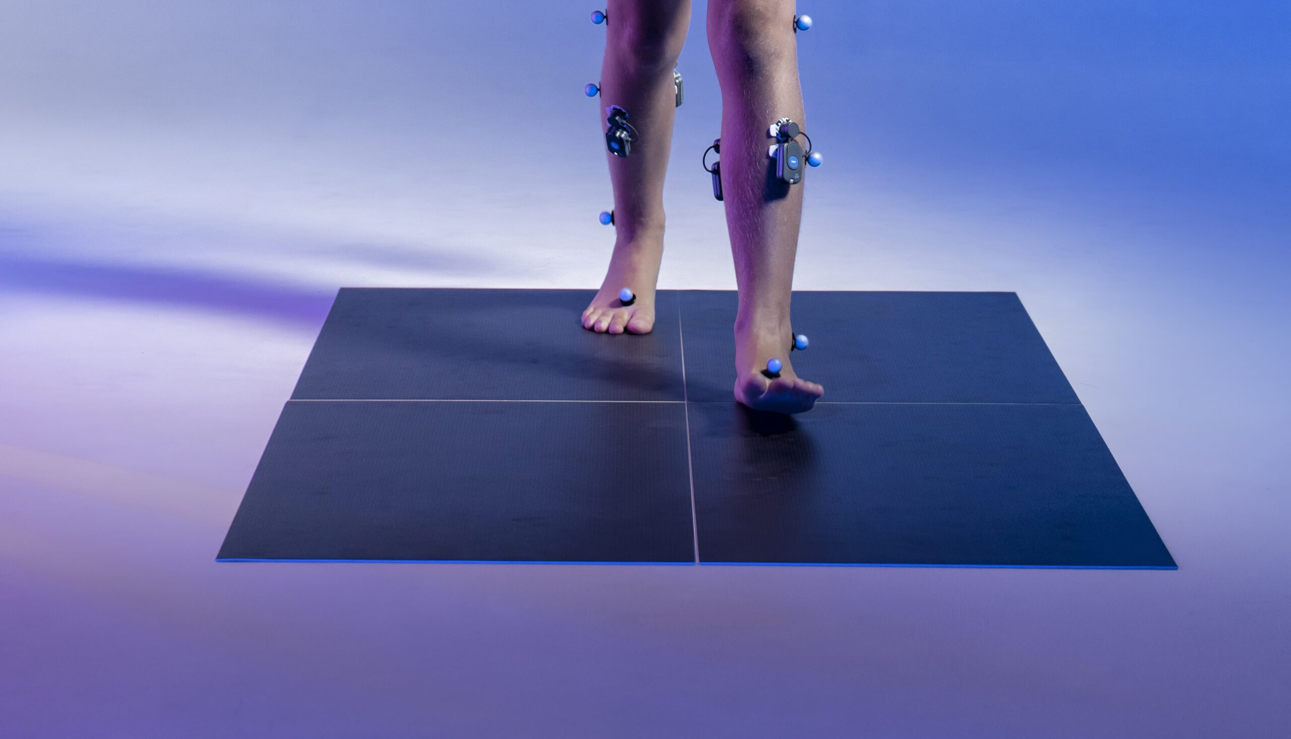
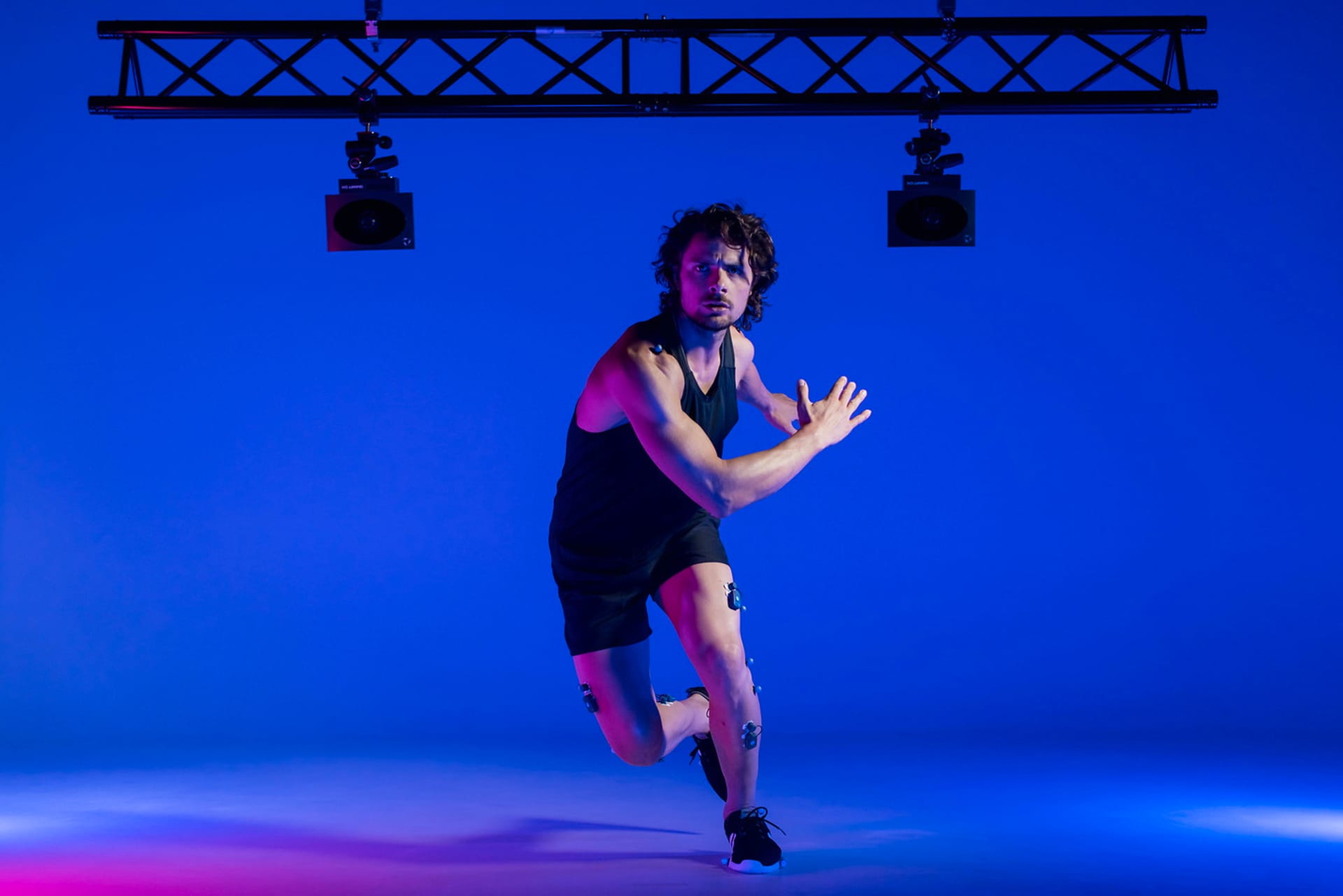
There was a need to further investigate the kinematics of the spine and whole body and to extend the conventional Helen Hayes M.M. protocol [6]. For this reason, a group of researchers at the University of Campania (Naples – Prof. P. De Blasiis) implemented a new protocol using a market set titled DB-Total protocol, with the aim of better assessing the sagittal spine and whole-body posture of healthy subjects and patients in the standing position. This protocol placed a total of 37 additional skin markers on the head, spine, and upper limbs compared to the Helen Hayes marker set [7].
The present study aims to analyze the new DB-Total parameters in a healthy adult population during walking to define their kinematic curves, additional values (mean of mean, range of motion, absolute range of motion), intra- and inter-subject repeatability.
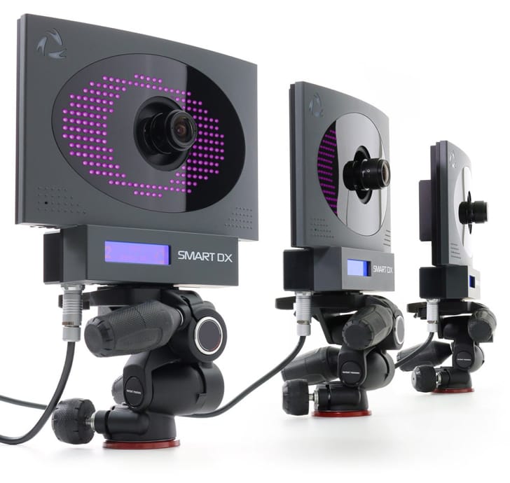
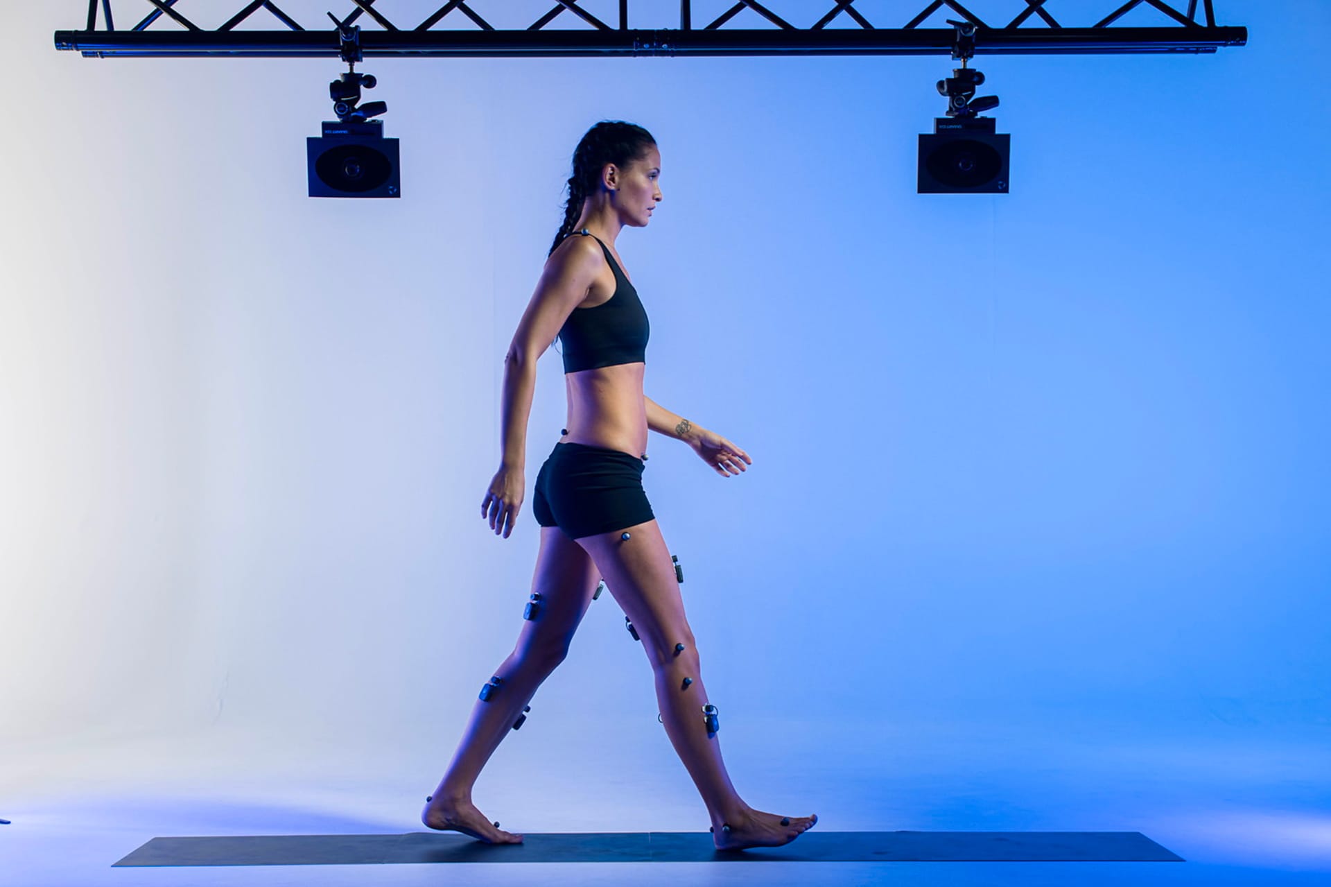
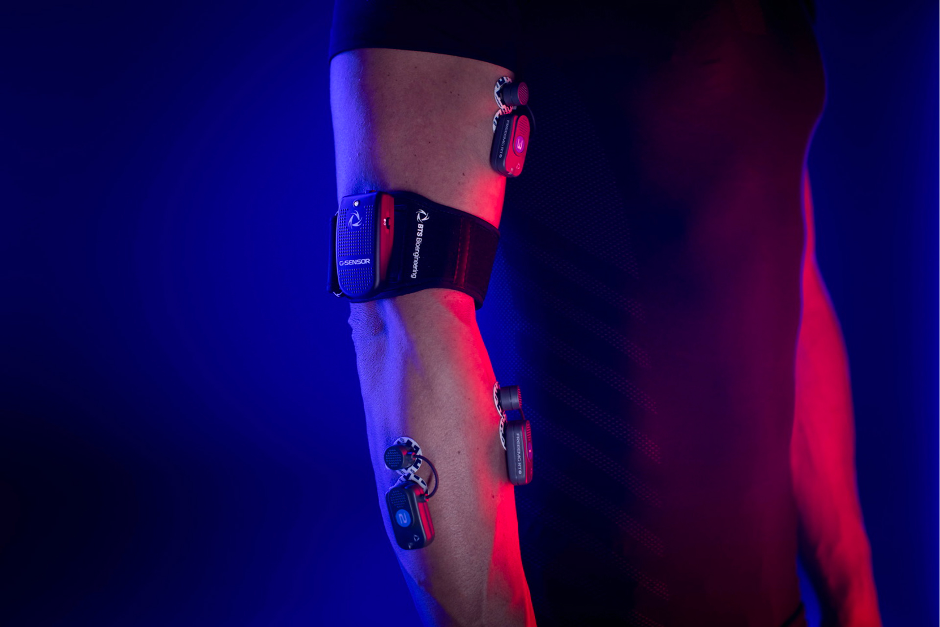
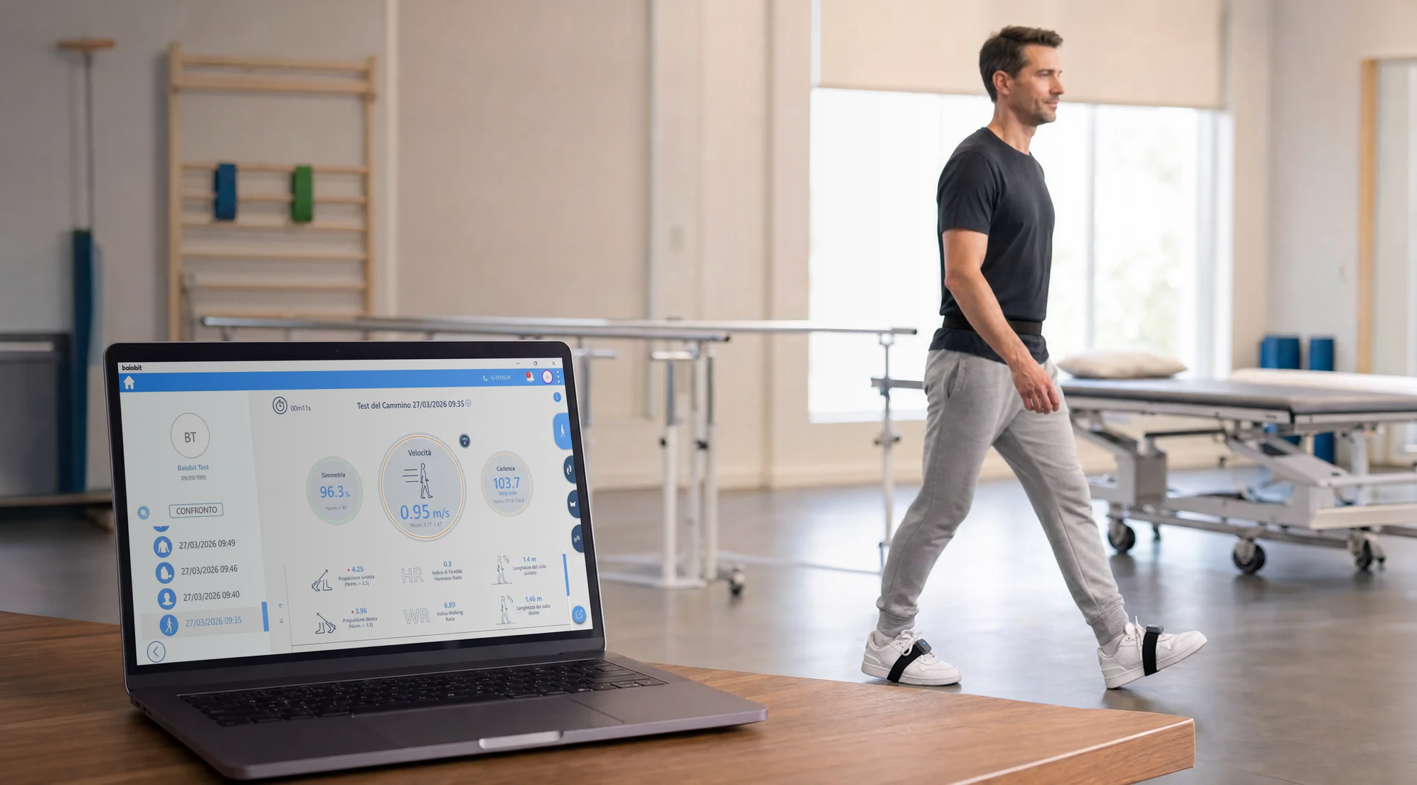
The research team conducted a preliminary cross-sectional observational study, recruiting fourteen healthy subjects. Each subject underwent a 3-D stereophotogrammetric examination, performed with 8 Smart-D optoelectronic cameras and 2 force platforms (BTS Bioengineering, Milan, Italy). Reflective markers were placed on body landmarks according to the new DB-Total marker set [8]. Each subject performed three consecutive trials in the same direction, walking barefoot on a 6-m walkway at a self-selected normal speed. The raw data were processed with Smart Analyzer software (BTS Bioengineering, Milan, Italy). In addition to conventional spatiotemporal parameters and conventional kinematic parameters, the DB-Total protocol allowed the calculation of 18 new kinematic parameters, including specific thoracic and lumbar angles and several vertical sagittal axes (Fig. 1).
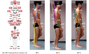
Fig.1 (A) DB-Total marker set protocol. (B) Graphic representation and definition of 18 sagittal whole-body parameters. Additional DB-Total markers were circled in red with respect to Helen Hayes Medial Marker set ones (De Blasiis et al, 2022)
The results of this novel method showed that the spatiotemporal parameters have very small SD and range of motion values (low inter-subject variability, CV<7%), except for stride speed, double stance phase and stride width. These results indicate good homogeneity of gait in all healthy subjects, which makes statistical analysis of kinematic parameters more reliable.
As for the new DB-Total parameters, the analysis showed low intra-subject variability (CV<50%) for most of them, which means high intra-subject repeatability of their kinematic curve during the gait cycle. In particular, these results showed a consistent pattern for the parameters of the more fixed (pelvis-dorsal) versus the more mobile (cervical spine-head-upper limb) body regions. One explanation could be related to the need to keep the heavier and more rigid body districts (pelvis and rib cage) within the support area between the two feet; therefore, the heel-T7/C7 segments should be aligned, while the more mobile ones (lower limbs, lumbar spine, and cervical spine) can adapt to movement.
The results of the study defined the kinematic curves and normal ranges of the new DB-Total parameters that analyzed, from the bottom up, the correlations between heel, pelvis, multi-segmental trunk, head and upper limb positions during different phases of the gait cycle. This protocol made it possible to measure additional whole-body kinematic parameters during walking, and could therefore help diagnose and better understand whole-body kinematic deviations in an adult pathological population.
References
- Ivanenko, Y.; Gurfinkel, V.S. Human Postural Control. Front. Neurosci. 2018, 12, 171.
- Fullin, A.; Caravaggi, P. Variability of postural stability and plantar pressure parameters in healthy subjects evaluated by baropodometry. Int. J. Environ. Res. Public Health 2022, 19, 2913.
- Moffa, S.; Perna, A. Effects of Hoverboard on Balance in Young Soccer Athletes. J. Funct. Morphol. Kinesiol. 2020, 5, 60.
- Leardini, A.; Sawacha, Z.; Paolini, G.; Ingrosso, S.; Nativo, R.; Benedetti, M.G. A new anatomically based protocol for gait analysis in children. Gait Posture 2007, 26, 560–571.
- Kinel, E.; D’Amico, M.; Roncoletta, P. Normative 3D opto-electronic stereo-photogrammetric sagittal alignment parameters in a young healthy adult population. PLoS ONE 2018, 13, e0203679
- Kadaba, M.P.; Ramakrishnan, H.K.; Wootten, M.E. Measurement of lower extremity kinematics during level walking. J. Orthop. Res. 1990, 8, 383–392.
- De Blasiis, P.; Fullin, A.; Sansone, M.; Perna, A.; Caravelli, S.; Mosca, M.; De Luca, A.; Lucariello, A. Kinematic Evaluation of the Sagittal Posture during Walking in Healthy Subjects by 3D Motion Analysis Using DB-Total Protocol. J. Funct. Morphol. Kinesiol. 2022, 7, 57.
- De Blasiis, P.; Fullin, A.; Sansone, M.; Del Viscovo, L.; Napolitano, F.; Terracciano, C.; Lus, G.; Melone, M.A.B.; Sampaolo, S. Quantitative Evaluation of Upright Posture by x-Ray and 3D Stereophotogrammetry with a New Marker Set Protocol in Late Onset Pompe Disease. J. Neuromuscul. Dis. 2021, 8, 979–988.
